THERE IS A DIFFERENCE BETWEEN A LABORATORY LEAK AND A CAREFULLY PLANNED CONSPIRACY TO KILL PEOPLE, GENOCIDE USING DEADLY NANO WEAPON.
Do you see the difference?
There are a growing number of researchers and scientists who are EXPOSING the fact that "Covid" IS NOT A VIRUS.
It is an OXIDATIVE STRESS, ACUTE OXIDATIVE STRESS, which wreaks havoc on the body, causes severe damage and ultimately death due to the toxicity of the nanotechnology used.
People have been given NANO POISON in drugs, PCR tests, masks, flu vaccines, chemtrails, food, water and of course C-19 injections.
What's more, they are given this NANOTECHNOLOGY under the pretext that it will protect them, for example, that it destroys bacteria and thus viruses. How does it destroy bacteria? It cuts through them!!!
https://pubmed.ncbi.nlm.nih.gov/20925398/ Toxicity of graphene and graphene oxide nanowalls against bacteria - PubMed (nih.gov)
On the basis of measuring the efflux of cytoplasmic materials of the bacteria, it was found that the cell membrane damage of the bacteria caused by direct contact of the bacteria with the extremely sharp edges of the nanowalls was the effective mechanism in the bacterial inactivation.
How does graphene damage viruses, bacteria and human cells? Graphene is a thin but strong and conductive two-dimensional sheet of carbon atoms. There are three ways that it can help prevent the spread of microbes: – Microscopic graphene particles have sharp edges that mechanically damage viruses and cells as they pass by them.
In one 2016 experiment, mice with graphene placed in their lungs experienced localized lung tissue damage, inflammation, formation of granulomas (where the body tries to wall off the graphene), and persistent lung injury, similar to what occurs when humans inhale asbestos. A different study from 2013 found that when human cells were bound to graphene, the cells were damaged.
One such study published in March 2020 found that a lifetime of industrial exposure to graphene induced inflammation and weakened the simulated lungs’ protective barrier.
Another study suggested that the irregular protrusions and sharp edges of the nanosheets could damage the plasma membrane, thus letting G entering the cell by piercing the phospholipid-bilayer (Li Y. et al., 2013):
https://www.pnas.org/doi/full/10.1073/pnas.1222276110 Graphene microsheets enter cells through spontaneous membrane penetration at edge asperities and corner sites | PNAS
Local piercing by these sharp protrusions initiates membrane propagation along the extended graphene edge and thus avoids the high energy barrier calculated in simple idealized MD simulations.
https://newatlas.com/graphene-bad-for-environment-toxic-for-humans/31851/
Two recent studies give us a less than rosy angle. In the first, a team of biologists, engineers and material scientists at Brown University examined graphene’s potential toxicity in human cells. They found that the jagged edges of graphene nanoparticles, super sharp and super strong, easily pierced through cell membranes in human lung, skin and immune cells, suggesting the potential to do serious damage in humans and other animals…
Those who did it play dumb.
First of all, they hide what exactly they administered and why.
https://www.sciencedirect.com/science/article/pii/S2352940718302853#fig0035 Investigation into the toxic effects of graphene nanopores on lung cancer cells and biological tissues - ScienceDirect
Since the isolation of graphene in 2004, research has been conducted to elucidate the potential toxic effects of graphene exposure in in vitro and in vivo environments. Much research has been carried out on pristine graphene, graphene oxide, reduced graphene oxide, graphene quantum dots, and graphene nanoribbons and has showed that these single or few-layered structures are capable of inducing adverse effects in cell lines and animal models [38], [39]. These early investigations initiated many pre-clinical toxicity studies on graphene nanostructures designed to inform the potential use of these structures in clinical settings. The results of these studies suggest that graphene nanostructures such as graphene oxide and reduced graphene oxide, have the capacity to induce toxicity in mammals both as a function of their chemistry, by inducing oxidative stress and lipid peroxidation, and as a result of their aggregation causing physical blockages [40]. Indeed, 3D porous graphene frameworks have shown various effects from acute lethally to sub lethal toxic effects including histological, and oxidative stress responses and, after inhalation exposure in rats, graphene has been found to accumulate in the lung, leading to phagocytosis [30].
However, GNPs, one of the most prominently used derivatives of graphene, e.g. used in DNA sequencing, drug delivery cargos and water treatment, have not been investigated for their potential toxicity [41].
They do not inform the victims what the antidote to the negative effects of this technology, this poison, is.
A comprehensive post mortem histological study was then performed to assess any tissue interactions with GNPs.
GNPs at all dosing regimens induced pathological changes after 27 days. Specifically, vacuolation, dilation of central vein and haemorrhage, vacuolation and dilation of central vein, damage of vacuolation, haemorrhage and degeneration of central vein, dilation of epithelial lining and hydropic degeneration oedema were observed in liver tissue. Kidney tissue of the treated groups showed acute vacuolization, dilation of epithelial lining, vacuolation and nucleus degeneration, nucleus damage, necrosis and epithelial degeneration. Heart tissue showed chemodectoma, toxic myocarditis, reddish brown atrophy; yellowish brown pigments suggesting lipofuscin granules as remnants of cell organelles and cytoplasmic material. The brain showed effects of secondary carcinoma, olegodendrocytoma small thin walled blood vessel and crytococcosis. Testicular tissue of treated groups showed spermatogenesis and vacuolation, dilation of germinal layer, degeneration of secondary spermatocytes, damage to the germinal layer and vacuolation. The lung showed damage of vacuolation, degeneration of central vein, inflammation, haemorrhage, d-shaped cells structure, hemosidophroages and lesion.
These nanostructures, depending on their composition and physiochemical properties can produce severe damage to cells by inducing oxidative stress [44]. An understanding of the toxicity mechanism is vital to attaining a more uniform understanding and comparison of observed effects.
Our data support previous studies that have demonstrated accumulation of graphene nanosheets in the
liver, lung, kidneys, and spleen
after intraperitoneal, intravenous, or dermal administration [49].
https://www.sciencedirect.com/science/article/abs/pii/S0009279723000637 Quantum dots are time bomb: Multiscale toxicological study - ScienceDirect
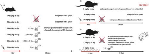
We found that QDs had a dose-dependent species-specific toxic effect on animals. At doses greater than 30.0 mg/kg,
mice died of spleen, kidney, and liver damage.
Over 48 hours, distribution was mainly observed to liver, adrenal glands, spleen and ovaries, with maximum concentrations observed at 8-48 hours post-dose.
So, yes, biodistribution, toxicity/adverse reaction studies after administration of these C-19 products, patents, shedding (https://outraged.substack.com/p/shedding), money invested, etc. all indicate that graphene/carbon-based materials are PURPOSELY being unleashed on the population.
The people behind this conspiracy also pretend, and will continue to pretend, that a higher necessity caused the use of these components and the implantation of TOXIC nanosensors in people without their knowledge, because, after all, it's all because of this deadly "virus".....
No, it is a pandemic of nanotechnology toxicity from beginning to end.
So, why were people hospitalized with pulmonary problems / flu-like symptoms and suffocation?
http://dol1.eng.sunysb.edu/pulled/nanotoxic-11032003-pdf.pdf
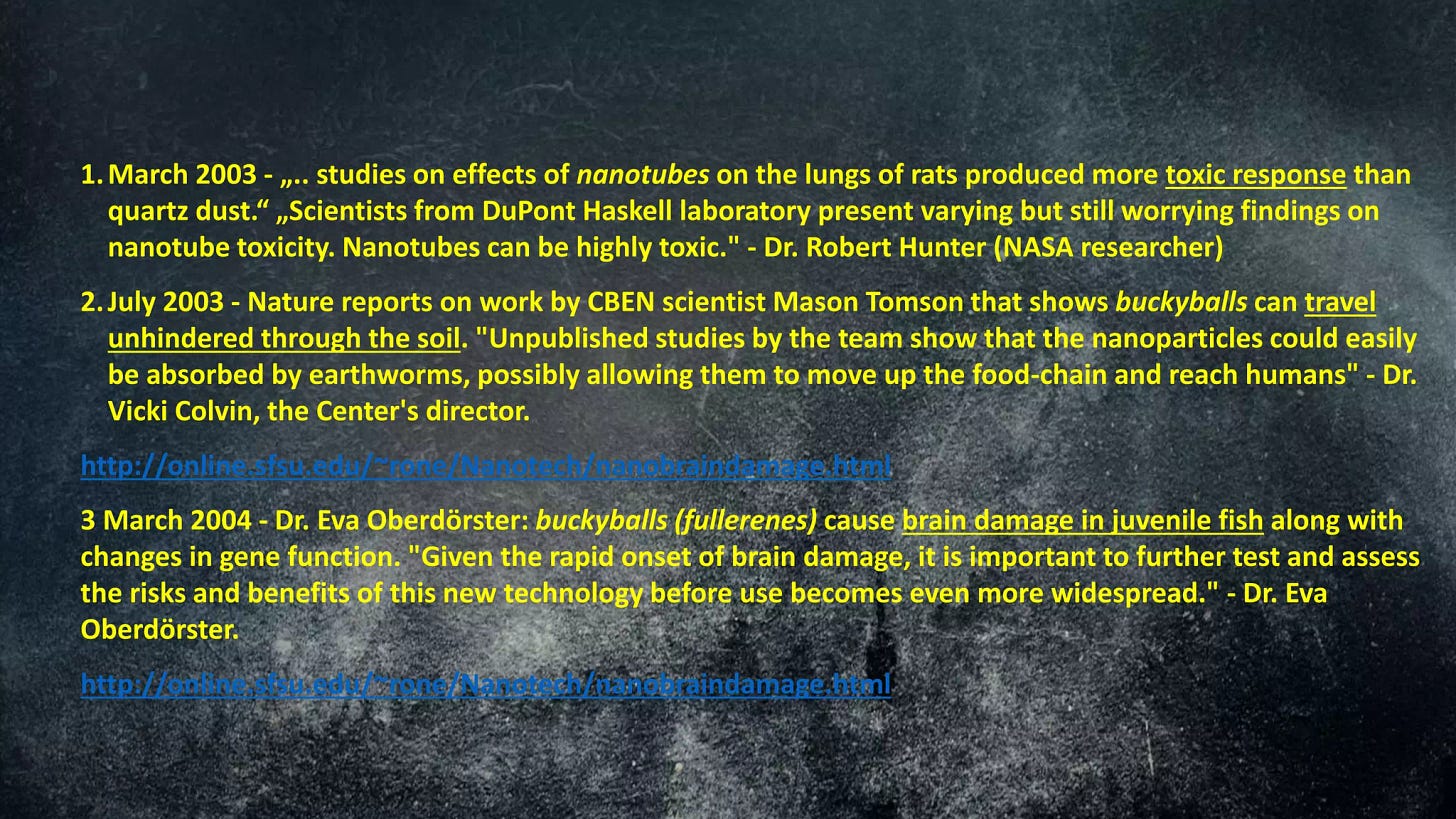
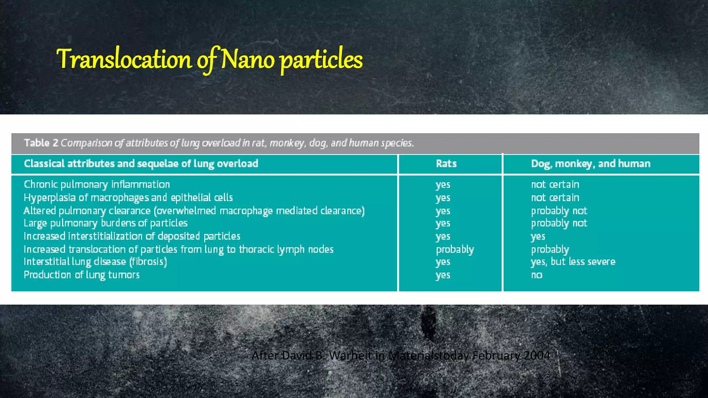
Take the experience of researchers at DuPont, who are testing microscopic tubes of carbon, known as nanotubes, valued for their extraordinary strength and electrical conductivity.
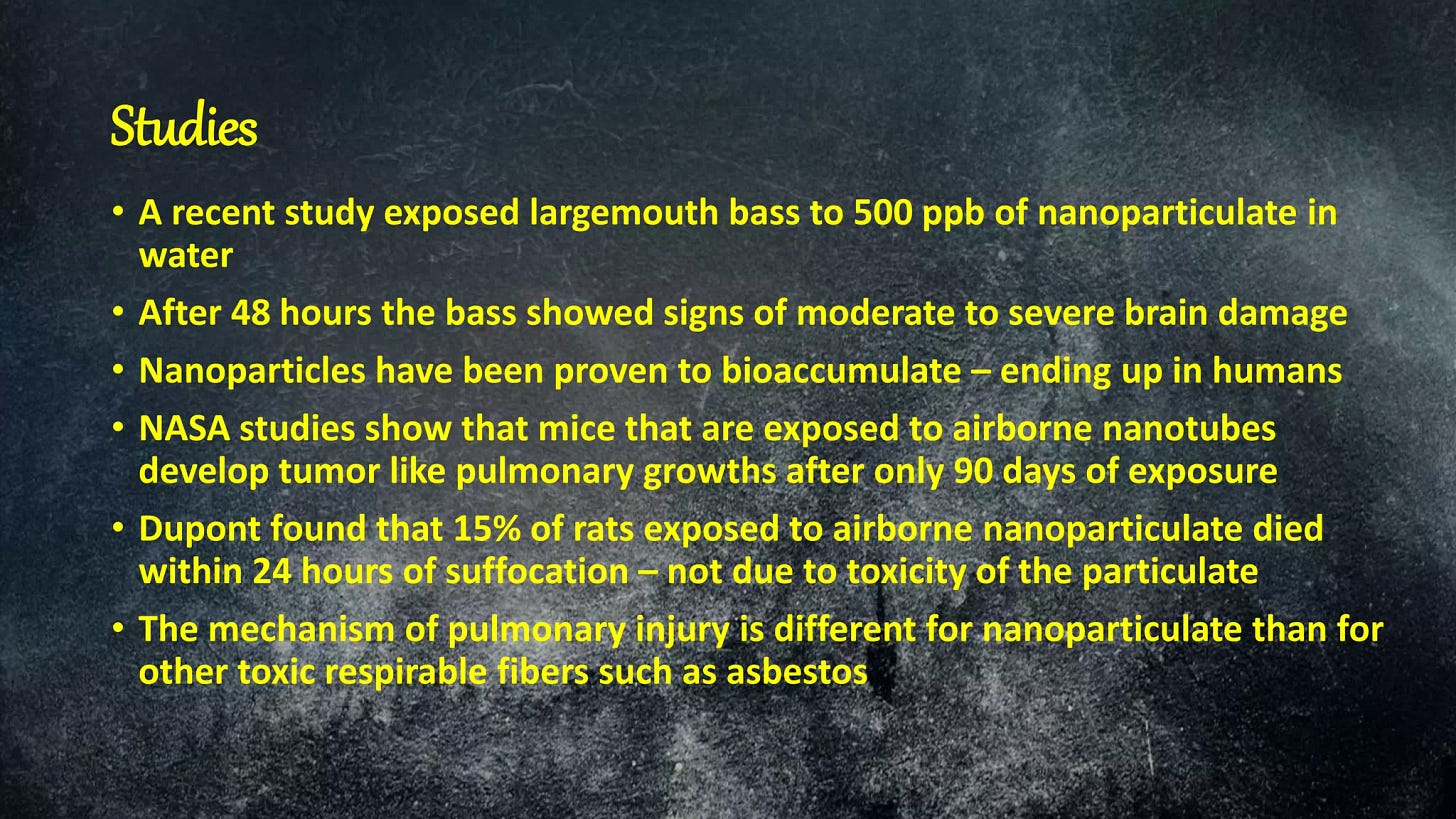
When the researchers injected nanotubes into the lungs of rats in the summer of 2002, the animals unexpectedly began gasping for breath. Fifteen percent of them quickly died. ''It was the highest death rate we had ever seen,''
said David B. Warheit, the research leader, who began his career studying asbestos and has been testing the pulmonary effects of various chemicals for DuPont since 1984.
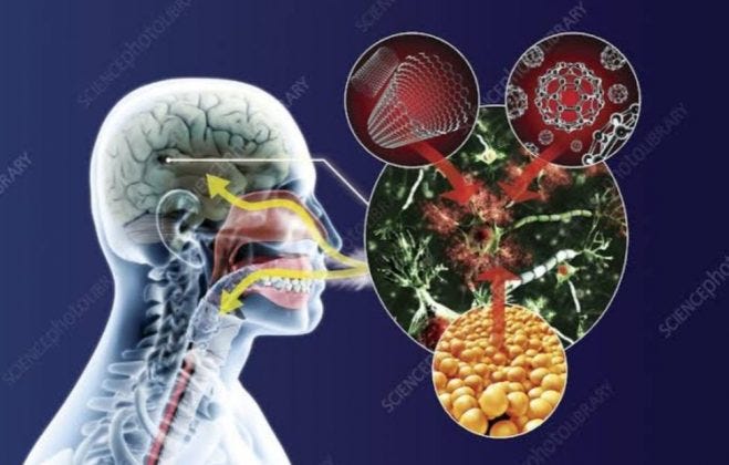
Lungs are not the only concern. Research shows that nanoparticles deposited in the nose can make their way directly into the brain. They can also change shape as they move from liquid solutions to the air, making it harder to draw general conclusions about their potential impact on living things.
https://www.ncbi.nlm.nih.gov/pmc/articles/PMC4706753/ INHALATION EXPOSURE TO CARBON NANOTUBES (CNT) AND CARBON NANOFIBERS (CNF): METHODOLOGY AND DOSIMETRY - PMC (nih.gov)
A high dose rate and high doses may overwhelm normal defense mechanisms and thus
result in significant initial pulmonary inflammation,
and may also affect disposition of the administered material to secondary organs.
Pulmonary exposure to SWCNT resulted in a rapid but transient inflammatory and injury response, as evidenced by increased levels of bronchoalveolar lavage fluid (BALF) neutrophils, lactate dehydrogenase (LDH) activity, and protein. Granulomas, predominantly in the terminal bronchioles, were reported 1 wk postexposure and persisted through 3 mo postexposure. A 15% rise in mortality rate within 1 d postexposure was noted and attributed to physical blockage of conducting airways by large SWCNT agglomerates.
https://pubmed.ncbi.nlm.nih.gov/15951334/ Unusual inflammatory and fibrogenic pulmonary responses to single-walled carbon nanotubes in mice - PubMed (nih.gov)
A rapid and transient inflammation and pulmonary damage were noted. In addition, granulomatous lesions and interstitial fibrosis within 7 d postexposure, which lasted through the 59-d course of the study, were observed. Granulomas were associated with the deposition of agglomerates in the terminal bronchioles and proximal alveoli, while interstitial fibrosis was associated with deposition of more dispersed SWCNT structures in the distal alveoli.
“Several studies have shown that carbon nanotubes cause lung cancer. The small tubes induce inflammation in a somewhat similar way to asbestos. Reprotoxic properties have also been observed. Up until now, the debate about the safety of nano has focused on the fact that more research is needed. However, here is a perfect example of where there is enough science to say that these materials should not be used”, says Dr. Lennquist.
https://www.ncbi.nlm.nih.gov/pmc/articles/PMC5468375/ Graphene and the Immune System: A Romance of Many Dimensions - PMC (nih.gov)
Of particular importance in this context is the fact that nano-biomaterials and pharmaceutical products alike are commonly contaminated with endotoxins which could lead to septic shock and organ failure if administered to patients (17).
Furthermore, modeling studies suggested that graphene, due to its hydrophobic nature,
may interrupt hydrophobic protein–protein interactions (35).
Indeed, it is important to recognize the differences in physicochemical properties between different members of the GBM family, not least with respect to the potential interaction with proteins.
Intravenously injected nanomaterials can adsorb a wide range of proteins in the blood (39). The bio-corona of blood proteins is rapidly formed, and it has been shown to affect hemolysis and thrombocyte activation (40).
Finally, in another recent study, nano-sized GO sheets were shown to induce
membrane ruffling in a variety of different cell lines with concomitant shedding of membrane fragments (65).
The underlying mechanism was not disclosed, although changes in the levels of Ca2+ in the cell are known to regulate the formation of such actin-driven membrane protrusions. Thus, it appears that GBMs are capable of interacting with cells in a variety of different ways including masking, piercing, ruffling/shedding, pore formation (possibly via membrane lipid extraction), and/or internalization into cells.
Taken together, a range of carbon-based nanomaterials including not only fiber-like materials but also spherical particles and flat materials such as GO trigger inflammasome activation.
And so it goes on...
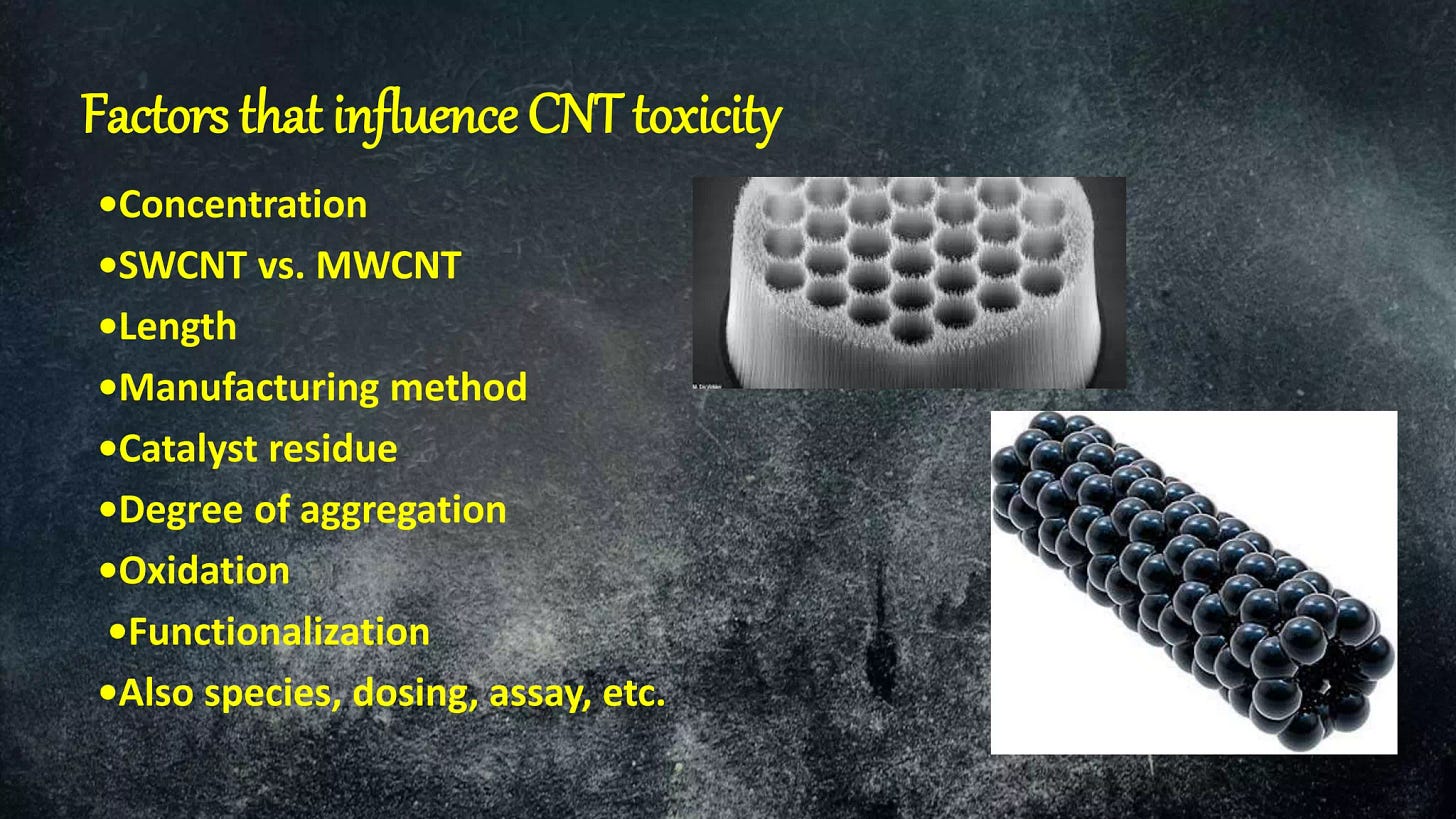
https://www.slideshare.net/19joyce/toxicity-of-fullerene-and-carbon-nano-tubes






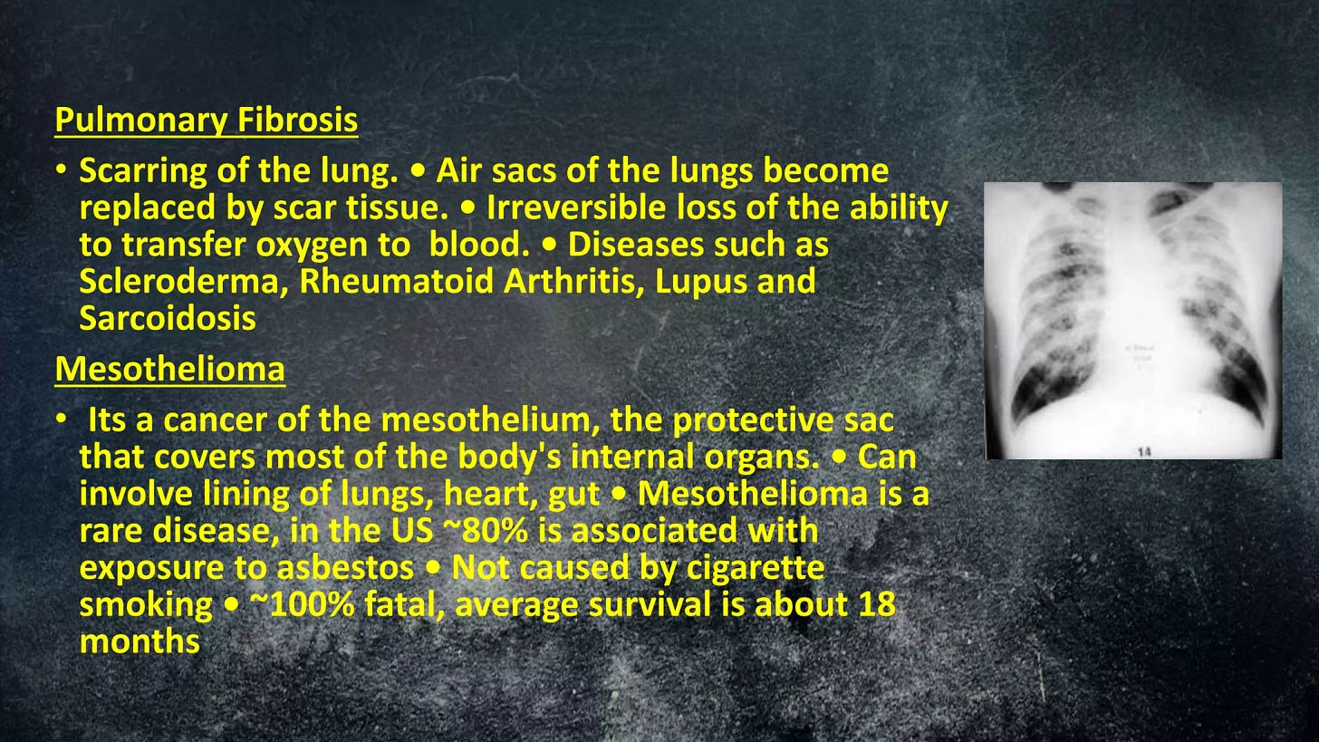

Infuriating and so hard to believe and bear that my husband took this and is gone! When will these people be punished???
Ah, I can agree on statements about the assault, and wish it weren’t just graphene, just this or that. It’s more complex than that, including molecular components and mechanisms in a majority of the global population already. The challenge is that all the easy going scientists who work on these components, are like a trio of people at an American style capital punishment event. One loads one chemical, another something else, while one directs the team and another pushes a lethal plunger. None feel responsible for killing that person strapped to the chair.
Plausible deniability in function, pace and order of damaging mechanisms is all as planned in leading countries’ plans for weaponization of manmade, man extracted, isolated and assembled natural molecules,
Sick? Killer of a well paid job by American military, it’s contractors, aspiring PhDs and lobbyists or political grifters... with no blame, shame or change in behavior.
Why? Not a single person to blame or liable for death. Murderous crowd - in the name of homeland security and patriotism.