WHAT'S THE DIFFERENCE BETWEEN C-19 AND ZEBOV INJECTION, DR. MALONE?

Sasha Latypova has just had an exchange with R. W. Malone:
https://sashalatypova.substack.com/p/dear-dr-malone
Robert Malone recently published a post revisiting his reasons for getting vaccinated with Moderna and his subsequent vaccine injury:
In early 2021, I spoke to colleagues at the FDA, who assured me that the vaccine was safe and was helping people with long COVID. They also re-assured me that the toxicity that I had seen with the LNPs in the 1990s had been resolved with new formulations. I trusted my government and public health. I trusted my friends. At that point, almost no one was writing about myocarditis or even that the vaccine was producing any adverse events. Frankly, I was proud of my part in inventing this suite of technologies and glad to see the ideas evolve into something that would save lives. Little did I know that all of the “research and development” in the years after I had left the field had never addressed that original safety issues and that in fact the addition of pseudouridine had made these products even more dangerous. Remember- most corporate research is never published - literally we the public have no way of knowing what had been done or not done. We had to trust the government.
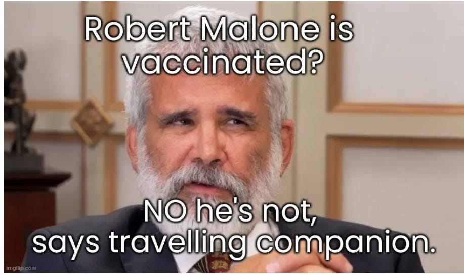
However, according to Steven Hatfill, who traveled to Italy with R.W. Malone, Robert Malone was never vaccinated:
https://anthonycolpo.substack.com/p/robert-malones-travelling-partner
“Curious then, that Hatfill recalls how he and Sly Malone had to submit to rapid antigen tests in Italy because they were both unvaccinated.
“I’m not vaccinated, I don’t have a card” says Hatfill.
“Neither did Dr Malone,” adds Hatfill with a big grin, “he didn’t have a card.”
My take on that? Of course, he never took it!
So why does he resort to such claims?
Well, looking at his contribution to the “Zika pandemic,” it seems that he is the creator of the current “countermeasures” as well…
https://web.archive.org/web/20170624121734/http://www.zikaresponse.org/news/

https://web.archive.org/web/20170623034210/http://www.zikaresponse.org/
History
The Zika Response Group began in the winter of 2016.
News of Zika was rapidly spreading and the founders of iORG, Dr's Robert and Jill Malone wanted to do something to really make a difference.
Having worked in infectious disease outbreak response teams for many years, they realized that the usual governmental response was slow and inefficient.
The usual response time just was not a good solution to solve the spread of Zika and to develop a better understanding of the disease, so that treatments could be developed.
Robert spoke to many colleagues and the response was overwhelming. together, they founded the Zika Response Group with iORG as the parent organization. The was goal was simply to impact on the public health response to make it more efficient and to make a difference. The team of people assembled worked night and day - and accomplished amazing things.
Some of the early successes are:
Zika Response Working Group wrote a comprehensive meta analysis of Zika and available medical countermeasure options that has helped guide government officials, clinicians and front line public health workers as they are responding to the virus outbreak in January 2016.
Zika Response Working Group wrote an extensive review paper on Zika that has helped guide scientists about the virus. Published in PLoS NTD. The paper now has over 40,000 readers and is one of the most cited Zika references.
Members of the Zika Response Group have performed epitope mapping and computational biology analyses, showing
the evolution of the virus - which will lead to a better vaccine
and a better understanding of how the disease is causing damage to fetuses, as well as the mechanisms of action of GBS. This effort culminated in two peer reviewed papers being published. The Evolution of the Zika Virus paper, published in the fall of 2016, is one of Wiley's most read scientific articles, and is in the 99% of their top publications- with 72,000 readers having already viewed the paper.
Members of our team have extensively consulted with USG and Brazilian government officials on how to control the pandemic.
Members of our team have consulting with the World Health Organization, and foreign public health officials on how to control the pandemic.
A member of the Zika Response Working Group is working in South America on GBS.
Members of our team have discovered possible anti-viral drugs that are safe for pregnant women and designed clinical trials, and are in the process of procuring funding and building partners in Latin America to move forward with clinical trials!
SAFE ANTI-VIRAL DRUGS??? DO YOU MEAN SMALL MOLECULE GS-5734 REMDESIVIR DRUG???
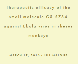
https://web.archive.org/web/20170625224344/http://www.nature.com/nature/journal/v531/n7594/full/nature17180.html Therapeutic efficacy of the small molecule GS-5734 against Ebola virus in rhesus monkeys
https://en.wikipedia.org/wiki/Remdesivir
Remdesivir (Veklury) is the international nonproprietary name (INN) while the development code name was GS-5734.
https://web.archive.org/web/20170623034210/http://www.zikaresponse.org/

Mission
The Zika Response Working Group, under the pending non-profit non-governmental organization iOrg (Infectious Outbreak Response Group), is rapidly responding to critical needs. Our activities are focused on;
· Threat and risk assessment and determination
· Identifying existing licensed drugs likely to have anti-Zika activity
· Modeling the outbreak to help inform government and corporate planning
· Geo-spatial tracking and sequence analysis
· Computational immunology and comparative proteome analysis including epitope mapping
· Using surveys and alternative media to track the spread of Zika virus, Zika disease, Zika communication, and to identify the unmet needs of medical caregivers and their patients
· Diagnostics and diagnostic technology research and development
· Development, manufacturing and production of medical countermeasures for Zika disease (devices, drugs, biologicals, and vaccines)
VACCINES??? DO YOU MEAN ZEBOV???

Contraindications and Precautions for Ebola Vaccine
The main contraindication for all three Ebola vaccines is
Severe allergic reaction (eg, anaphylaxis) to any component of the vaccine
Precautions for all 3 Ebola vaccines are
Anaphylaxis
Thrombocytopenia or any coagulation disorder
Anaphylaxis has occurred after administration of these vaccines.
After vaccination, people should be observed for at least 15 minutes for early signs of anaphylaxis or anaphylactoid reactions.
👆👆👆👆❗❗❗❗❗👆👆👆👆❗❗❗❗❗👆👆👆👆❗❗❗❗❗
Appropriate medical treatment and supervision must be available in the event anaphylaxis occurs.
The vaccines should be given with caution to people with thrombocytopenia or any coagulation disorder because bleeding or bruising may occur after the vaccines are given.
Adverse Effects of Ebola Vaccine
For all 3 vaccines, the most common adverse effects are injection site pain and swelling, myalgias, arthralgias, headache, and fatigue.
For rVSV-ZEBOV, other common adverse effects are injection site redness, feverishness, nausea, arthritis, rash, and abnormal sweating.
For Ad26.ZEBOV/MVA-BN-Filo, other common adverse effects include injection site warmth and chills. In children 1 to 17 years of age, the most common adverse effects reported are”Injection site pain
Fatigue
Decreased activity
Decreased appetite
Irritability
The rVSV-ZEBOV trial is funded by WHO, with support from the Wellcome Trust; the UK Government through the Department for International Development; the Norwegian Ministry of Foreign Affairs; the Norwegian Institute of Public Health through the Research Council of Norway,; the Canadian Government through the Public Health Agency of Canada, Canadian Institutes of Health Research, the International Development Research Centre, and the Department of Foreign Affairs, Trade and Development; and Médecins Sans Frontières.
AND HOW ABOUT THIS?
https://www.medscape.com/viewarticle/821223_7?form=fpf

Abstract and Introduction
Abstract
siRNAs have immense therapeutic potential for the treatment of various gene-related diseases ranging from cancer, viral infections and neuropathy to autoimmune diseases. However, their bench-to-bedside translation in recent years has faced several challenges, with inefficient siRNA delivery being one of the most frequently encountered issues.
In order to improve the siRNA delivery especially for systemic treatment, nanocarriers made of polymers, lipids or inorganic materials have become almost essential.
The 'negative' aspects of these carriers such as their nanotoxicity and immunogenicity thus can no longer be overlooked.
In this article, we will extensively review the nanotoxicity of siRNA carriers.
The strategies for mitigating the risks of nanotoxicity and the methodology for evaluating these strategies will also be discussed. By addressing this often overlooked but important issue, it will help clear the way for siRNAs to fulfill their promise as a versatile class of therapeutic agents.
The clinical translation of siRNA therapeutics, however, has turned out to be more challenging, with inefficient siRNA delivery and the issues associated with the siRNA delivery vehicles being the key problems.[6,7]
Numerous nanocarrier systems have been developed to improve siRNA delivery (see reviews by Zhao et al. and Kesharwani et al.[12,13]). In this review, our focus is on the toxicity of these nanocarriers.
It should be noted that even though certain siRNA therapeutics are designed with the intent to be 'toxic' to specific target cells (e.g., anticancer effects in cancer treatment), one should distinguish between their efficacy (intended effects) and toxicity (undesirable effects).
As will be discussed, the toxicity of nanomaterials is often less target specific and more complex and unpredictable, and thus should be kept at a minimum level.
It is our hope that this review will help nanomedicine researchers and clinicians to be more aware of these carrier toxicity issues so safer therapeutic siRNA products with higher translational success can be developed.
Besides using siRNA conjugates,[18,19] the most common approach to improve siRNA delivery is the use of nanocarriers.[12,13]
Table 2 shows that all 15 trials for systemic treatment involved a nanocarrier, most notably the lipid nanoparticle (LNP) system (formerly named stabilized nucleic acid lipid particles).[20,204]
Considering that it is a common practice to use nanocarriers, it is impossible to overlook their toxicity when designing a systemic siRNA therapy.
Once downsized to nanoscale, many normally 'inert' materials can become substantially more reactive probably owing to the dramatic increase in total surface area.
This results in more extensive interactions between these materials and the biological systems, causing damage to human body at the organ, tissue and cell levels, and the manifestation of 'nanotoxicity'.[21]
siRNA nanocarriers deserve particular attention owing to their complexity and frequent inclusion of toxic cationic materials that tend to interact well with various biological components.[12] The transient nature of their RNAi activity (3 days to 1 week) also implies that frequent, repeated administration will be required for chronic diseases, and the risk of cumulative toxicity is expected to increased.[22]
Nanotoxicity can be accentuated by many factors (Table 3).
Our early understanding of these factors mostly came from findings related to environmental exposure to inorganic or simple organic particulate matters.[21]
For instance, the slim shape of an asbestos fiber allows this material to physically interfere with the chromosomes, disrupting normal cell activities, such as mitosis.[23]
Studies on carbon and titanium oxide particles indicated that the smaller their size the higher is the pulmonary toxicity they cause is.[24]
Schaeublin et al.'s study with gold nanoparticles showed that surface charge was also a key determinant of their toxic effects on cellular processes.[25]
Neutral nanoparticles caused cell death through necrosis in HaCaT cells, whereas charged nanoparticles induced apoptotic cell death.
Other factors such as nanocarrier ingredients, dose level and duration of exposure, and routes of exposure may also affect the toxicity of a nanocarrier.[21,26,27]
How do siRNA Carriers Cause Nanotoxicity?
siRNA nanocarriers for systemic delivery are developed to encapsulate siRNAs, stay in the circulation, deliver the siRNA payload to the target cells, interact with the cell surface, enter the cell and efficiently escape the endosome–lysosome system to unload the siRNAs to the cytoplasm.[12,13]
To efficiently perform this series of tasks, researchers have introduced features to the nanocarriers that include surface modification with PEG (i.e., 'PEGlyated') to extend their circulation time,[28] controlled siRNA release optimization,[29,30] inclusion of cationic materials or targeting moieties to improve carrier–cell interactions,[31,32] addition of ingredients to enhance endosomal escape[33] or a combination of all of these.[13,34]
From a toxicological perspective, the high complexity of nanocarriers can be counterproductive. Each additional feature implies an extra risk of toxicity.
Table 4 summarizes the ways in which high levels of these complex nanodevices can lead to toxicity. Most siRNA carriers contain cationic ingredient(s) because cationic functional groups enable electrostatic complexation and better encapsulation of the polyanionic siRNA molecules.
The positive charges can facilitate interaction with the negatively charged cell surface.[35,36]
Cellular damage can be caused by direct interactions between the cationic groups and cellular components, or indirectly by reactive oxidative species (ROS) formed in the presence of cationic materials.
The resulting cell toxicity can be manifested in an acute or delayed manner.[37]
It should be noted that the mechanism about how cationic nanomaterials cause cytotoxicity is still not well understood and an alternative mechanism has been proposed.[38] Other obvious damage includes 'lysosomal overload' of poorly biodegradable nanocarriers, which results in the accumulation of visible autophagic vacuoles and apoptotic cell death.[39]
In addition to cationic materials,
production of ROS, such as hydrogen peroxide, superoxide anions and hydroxyl radicals, can also be triggered by noncationic nanomaterials (e.g., carbon nanotubes [CNTs] and metallic nanocarriers) or impurities (e.g., heavy metals) in the nanocarriers.[40,41]
Since the high surface area of nanomaterials tends to promote ROS formation, it has been suggested that
oxidative stress is a particularly significant mechanism responsible for nanotoxicity at the cellular level.[40]
High levels of intracellular ROS can react with a diversity of cellular macromolecules including DNA, proteins and membrane lipids. Based on the studies of CNTs, this could activate a number of molecular signaling pathways, including AP-1, and NF-κB and MAPK, which leads to the release of proinflammatory cytokines together with the depletion of antioxidant defenses, such as PARP1, p38 and serine–threonine kinase (Akt).
The risk of cell death is therefore increased.[42]
Many siRNA nanocarriers, not just CNTs, can stimulate the immune system via Toll-like receptor activation.[43] Secretion of cytokines and chemokines (e.g., TNF-α, IL-1β, IL-6, IL-10 and MCP1) can promote local inflammation. At in vivo and clinical levels this may recruit neutrophils that secrete additional proinflammatory cytokines (e.g., TNF-α and IL-1β). Other related cells such as macrophages and lymphocytes may also be attracted to the sight of inflammation.
While the immunostimulatory phenomenon can be exploited for therapy, more often it
may lead to undesirable outcomes such as incompatibility of the blood and the nanocarriers, hypersensitivity reactions andinflammatory responses and anaphylaxis due to suppressed immune defense.[43]
Excessive inflammatory responses can inflict significant damages to tissues and organs (e.g., liver toxicity) and can be lethal.
Nanocarrier-mediated toxicity can also occur in more obscure manners. At the cell level, nanocarriers can alter gene expression, a phenomenon known as toxicogenomics, and subtly disturb normal cell functions and phenotypes in a detrimental manner.[44]
It should be noted that severe disturbance of the expression of key proteins (e.g., p53, Rad51 and OGG1) can eventually cause cell mortality and noticeable tissue damage. Localization of very small nanocarriers in the cell nucleus can also induce DNA strand breaks (i.e., genotoxicity), and trigger cell death if it is excessive.[40]

Nanotoxicity of the Major Classes of siRNA Carriers
Cationic charges in siRNA carriers can be provided by various materials, most typically polymers, lipids and inorganic nanomaterials.
Polymer- & Dendrimer-based Nanocarriers
Polymers such as polyethylenimine (PEI), poly-L-lysine, poly-L-arginine, chitosan and their derivatives have been commonly used for siRNA nanocarrier development.[45] In addition, a class of highly branched, symmetric polymer-like macromolecules of 5–20 nm in size, known as dendrimers, have recently gained popularity for nucleic acid delivery.[45] Examples of dendrimers used for siRNA delivery include polypropylenimine (PPI) and polyamidoamines. These polymers and dendrimers have multiple cationic groups to facilitate complexation with siRNAs and enhance their endosomal escape using the proton-sponge effect.[45,46]
Among the cationic polymers, PEI has been the standard for nucleic acid delivery owing to its good cell membrane interaction, high cellular uptake rate and efficient endosomal escape.[47] PEI is available in several molecular weight grades and in branched or linear structures, with 25-kDa branched PEI being traditionally used most for siRNA delivery.[45] However, PEIs generally exhibit significant in vitro and in vivo toxicity.
Free, uncomplexed PEI molecules
can interact with negatively charged serum proteins and even red blood cell surfaces, to form aggregates to adhere to tissue surfaces to inflict acute cell damage.
PEI molecules can also be released from polyplexes internalized by cells and interact with cellular components to cause delayed toxicity.[48]
High-molecular-weight PEIs are more toxic. PEIs below 10 kDa generally have lower toxicity.[49] Branched PEIs often cause more cellular toxicity than the linear ones with similar molecular weights.[45,50] It was suggested that this is partly due to the higher efficacy of the branched PEIs compared with their linear counterparts. For instance, a study comparing branched PEIs with linear PEIs of the same molecular weight showed that branched PEIs induced stronger apoptotic responses in A431 cells both at early and late stage, resulting in more DNA damage and Akt kinase induction.[51] The only major difference between the two PEIs was that the branched form was more internalized by the cells. Similar findings were reported in nondifferentiated fibroblast COS-1 cells.[52]
However, other studies on PEI/DNA polyplexes showed that linear PEIs actually have both superior transfection efficiency and toxicity profiles.[50,53] The contradictory findings are probably attributable to the differences in the experimental conditions (e.g., in vitro vs in vivo) and the manner in which the polyplexes were prepared. The polyplexes formed by linear PEIs under salt-free conditions were found to be highly efficacious in vivo. Another report showed that while the proinflammatory activities of PEIs were independent of their structure, linear PEIs could be prepared at a higher N:P ratio (up to 25) to achieve improved in vivo efficacy, whereas branched PEI became lethal to the animals when the N:P ratio was higher than 15. Therefore, that linear PEIs will be more suitable for clinical use.
PEI toxicity is partly due to its limited biodegradability.[37,45] Hydrolysable analogous polymers with degradation half-lives in days, such as poly(amino esters), have, therefore, been developed.[54] It was shown that poly(amino ester) carrier achieved nearly a 1.5-times better gene-silencing effect and significantly lower cytotoxicity than 25-kDa PEIs. Other hydrolysable polymers such as poly(ester amine) and poly(amido amine) were also studied. Tzeng et al. tested them on human umbilical vein endothelial cells and demonstrated a 60–75% siRNA-mediated knockdown of GFP with high cell viability.[55]
Currently, the most successful synthetic cationic polymer used for siRNA delivery is probably a linear cyclodextrin-based polymer containing polycation (CDP).[56,57] In a nonhuman primate study, cynomolgus monkeys were systemically administered with CDP containing 3, 9 or 27 mg siRNA/kg.[57] Only mild elevations levels of the indicators of kidney toxicity (blood urea nitrogen and creatinine) and liver toxicity (ALT or AST) were observed at the highest dose level. Increases in IL-6 level in all animals and IFN-α in one animal was also detected, which is indicative of a modest immune response to the treatment. Meanwhile, there were no clinical signs of toxicity, changes in complement factors and hypersensitivity detected. The data indicated that except for a slight risk of dose-dependent toxicity in vivo, systemic use of CDP-siRNAs is safe. These favorable findings have propelled CDP-siRNA drugs onto the clinical stage.[58] In a Phase I trial in patients with solid refractory cancers, only grade 1–2 fatigue, fever/chills and gastrointestinal symptoms were observed at all dose levels, and transient increases in IL-6 and TNF-α were reported at the highest dose level.[59] Overall, this siRNA nanoformulation appeared to be well tolerated.
The concern for toxicity of synthetic polymers has led some researchers to explore other options, such as natural polymers and dendrimers. Chitosan, a naturally occurring polysaccharide composed of glucosamine and N-acetylglucosamine residues derived from the partial deacetylation of chitin, has been particularly studied owing to its low cell toxicity and immunogenicity and the polymer and chitosan/siRNA complexes.[60,61]'
Lipid-based nanocarriers
The cell membrane is rich in lipids and phospholipids, therefore, it is a logical choice to use lipids as materials to build nucleic acid delivery systems that demand good interaction with the cell surface. Cationic lipids have been widely used for DNA transfection for decades,[37,72] with N-[1-(2,3-dioleoyloxy)]-N-N-N trimethyl ammonium propane (DOTAP) as the most popular choice.[73] After the discovery of RNAi, there has been a general shift of RNAi application from DNA transfection to siRNA delivery. Currently, most of the products commercially available for siRNA transfection, for example, the Lipofectamine® series and Oligofectamine™ from Invitrogen (CA, USA) and RNAifect from Qiagen (Limberg, The Netherlands) are all cationic lipid based.[44]
Unlike polymeric nanocarriers, cationic lipid-based systems often include other noncationic ingredients, for example, neutral lipids. L-alpha dioleoyl phosphatidyl ethanolamine and cholesterol are frequently included to destabilize endosomes for endosomal escape and stabilize the nanocarrier, respectively.[73,74] These neutral lipids by themselves are generally less harmful than the cationic components, but may potentiate the overall carrier cytotoxicity in certain formulations.[75] It was reported that 293A cell viability was reduced from over 80% to below 60% when cholesterol was added to a DOTAP nanocarrier.
Hence, these apparently harmless ingredients should not be overlooked when considering carrier toxicity.
Most cationic lipid molecules consist of three regions: a cationic head, a hydrophobic hydrocarbon backbone and a linker that connects the head and tail.[73] The whole molecule resembles a detergent, therefore, a high concentration of cationic lipids
can compromise the membrane integrity resulting in cell lysis and necrotic death.
At a sublethal concentration, the lipoplexes can still cause irritation to the cells and induce cell shrinkage, vacuolization of the cytoplasm and a reduced number of mitoses.[76]
The cationic head groups are also capable of interacting with enzymes, such as PKC, to cause cell toxicity.[77]
Similar to cationic polymers,
cationic lipids also induce undesirable gene expression changes in vitro.
It was shown that Oligofectamine formulation upregulated expression of genes involved in the apoptotic process including caspase 8 isoform c, BCL2A1, HSP70 and HSPD1.
The authors suggested that the altered gene expression may lead to an increased tendency towards early apoptosis of the cells.
The preclinical findings on DNA lipoplexes are of some reference value on their in vivo toxicity as siRNA nanocarriers.
It was shown that after systemic administration of cationic lipid nanocarriers to animals, mortality occured at high doses.[81]
Systemic toxicities mediated by lipoplexes include:
Inflammatory toxicity: lipoplexes are captured by Kupffer cells and trigger the release of proinflammatory cytokines, such as TNF-α, IFN-γ, IL-6 and IL-12;
Liver toxicity: damage to liver tissues are indicated by elevated serum levels of transaminases especially ALT and AST;
Hematologic and serologic toxicity; for example, leukopenia and thrombocytopenia.[82]
CNTs are helical microtubules of graphitic carbon with outstanding stability and strength-to-weight and aspect ratios.[94] Several CNT-based systems have been developed for siRNA delivery[95] and they generally demonstrated a high delivery efficiency. However, in the past few years their toxicity has been under serious scrutiny.[96] The key issue surrounds their resemblance to asbestos fibers in terms of the high aspect ratio and, hence, pathogenic effects.[97] After intraperitoneal administration, the CNT molecules recruited inflammatory cells and caused thickening of mesothelioma in the lining of abdominal cavity. Needle-like CNTs and asbestos both also activated IL-1 secretion from primed macrophages.[98] It was found that the activation of NLRP3 inflammasomes essential for CNT-induced IL-1 secretion depended on harmful ROS production, CTSB activity, P2X receptor, and Src and Syk tyrosine kinases. Owing to these risks, caution should be exercised when choosing CNTs for siRNA delivery. The CNTs of a longer length should be avoided.
…
OF COURSE ROBERT MALONE KNOWS THIS VERY WELL, AS DOES HIS WIFE. HOWEVER, THEY ARE SILENT ON THE SUBJECT OF NANOTECHNOLOGY.
AND HOW ABOUT THIS?
No proof Zika causes microcephaly, UW-Madison study says
David Wahlberg | Wisconsin State Journal Dec 28, 2017
A new study by a UW-Madison professor calls into question the link between the condition and the Zika virus.
SO, WHO'S REALLY DR. MALONE…?
HERE IS MORE INFORMATION FROM THEM (https://web.archive.org/web/20160822020856/http://www.zikaresponse.org/news/) ON THESE MONSTROUS EXPERIMENTS CONDUCTED ON VICTIMS OF "ZIKA"
https://web.archive.org/web/20160822020856/http://www.zikaresponse.org/news/
"WHO may be leading Brazil down wrong path on Zika virus"
"Nearly a year after Zika began to be reported here, and five months after the government declared a public-health emergency, no one has yet found proof that Aedes aegypti is spreading Zika or whether it may be one among several vectors. One critical question is whether the virus can be transmitted by another mosquito – possibly one in the genus Culex, which is vastly more common than Aedes aegypti.
Yet Brazil, on the advice of the World Health Organization, has based its entire response to the Zika crisis on combatting this one mosquito species. And the political and economic crisis gripping the country has sidelined an already haphazard and underfunded response.
But Ms. Rousseff was wrong. Her government does not know even this one fact about Zika. Although there is good cause to suspect that Aedes aegypti, the mosquito species with distinctive black-and-white striped legs, is the agent of infection, there are no confirmed findings of the virus in aegypti in Brazil.
Nearly a year after Zika began to be reported here, and five months after the government declared a public-health emergency, no one has yet found proof that Aedes aegypti is spreading Zika or whether it may be one among several vectors. One critical question is whether the virus can be transmitted by another mosquito – possibly one in the genus Culex, which is vastly more common than Aedes aegypti.
"Scientists outside Brazil increasingly share her fears. Fiona Hunter, an entomologist with Brock University in St. Catharines, Ont., who attended the Aedes summit, said she is deeply concerned that the country might be making a mistake in its response. “The fact that there are no data as far as I can tell that Aedes aegypti is driving this Zika epidemic just flabbergasts me.”
Dr. Hunter was involved in the first surveillance to find the vector for West Nile in Canada. To do that, she said, scientists set traps to gather every mosquito existent in an area, then tested and retested dozens of species to establish beyond doubt which one was driving the epidemic.
“That’s how research into a new emerging disease ought to be done,” she said, and the vicious behaviour of Zika in Brazil merits treating it as a new disease. “But they didn’t figure it out – the Brazilians automatically assumed it’s aegypti. I hear physicians here on the radio in Canada saying you don’t have to worry ever about Zika – well, yes you do. There are implications for the spread of this virus around the world.”
Dr. Capurro in Sao Paulo said that she would like to be analyzing 2,000 mosquitoes a month but is constrained by the lack of a national network. “I need connections with hospitals [to identify prospective capture sites] and municipal administrations – who do I even call?” she said.
Larvicide Automatic Dispenser wins most Implementable Solution Award; Congrats to Adriano De Bernardo at UNC Charlotte and member of the Zika Response Working Group!
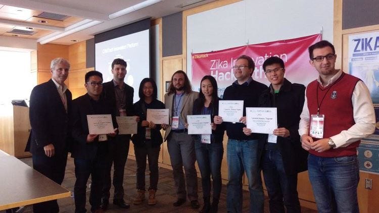
OUR OWN ADRIANO DE BERNARDI SCHNEIDER, MEMBER OF THE ZIKA RESPONSE WORKING GROUP AND PHD CANDIDATE, DEPARTMENT OF BIOINFORMATICS AND GENOMICS AT UNC CHARLOTTE WAS ON THE WINNING TEAM! THEIR PROJECT, FOR A LARVICIDE AUTOMATIC DISPENSER WON THE MOST IMPLEMENTABLE SOLUTION AWARD AT THE ZIKA INNOVATION HACK-A-THON THIS WEEKEND!
The event was organized by CAMTech Consortium for Affordable Medical Technologies, Global Disarter Response, Massachusetts General Hospital - Global Heath, MIT Hacking Medicine, Harvard, Global Health Institute, Design That Matters, Medtech Boston. The Hack-a-thon received generous sponsorship by GE Foundation, Johnson & Johnson, and Merck.
Their project, called L.A.D. (Larvicide Automated Dispenser), was awarded "Most Implementable Solution" sponsored by GE Foundation. The device has the potencial to help control mosquitos borne diseases such as Zika, Chikungunya, Dengue, and Malaria at the same time it reduces health care costs in countries affected by them.
A TIMELINE OF THE ZIKA RESPONSE WORKING GROUP'S ACCOMPLISHMENTS AND CONTRIBUTIONS TO THE FIGHT AGAINST THE ZIKA VIRUS.
BELOW IS A TIMELINE OF OUR ACCOMPLISHMENTS AND CONTRIBUTIONS TO THE FIGHT AGAINST THE ZIKA VIRUS. THE ZIKA RESPONSE GROUP, GOING AT THIS 24/7 HAS DONE AMAZING WORK AND HAVE MADE A DIFFERENCE: AND MOST OF IT HAS BEEN A VOLUNTEER EFFORT. THE GOOD NEWS IS THAT WE AREN'T FINISHED YET- SO KEEP CHECKING BACK FOR MORE DISCOVERIES, INSIGHTS AND MAYBE EVEN FURTHER DEVELOPMENT OF AN ANTI-VIRAL DRUG THAT COULD SAVE LIVES!
The timeline:
Zika Response Working Group have produced a white paper, that was submitted in January to Government Officials at the highest levels.
A member of the group, Dr. Robert Malone has discovered a class of drugs that act as anti-virals against Zika Virus, and which are safe for pregnant women. The World Health Organization has advocated these drugs to be developed as a first defense for pregnant women!
For more information, please contact Dr. Malone directly at rwmalonemd@gmail.com or go to www.atheric.com (a company set up to develop these anti-viral drugs).
A member of the Zika Response Working Group, Dr. Malone participated in a consultation in Genevea at the World Health Organization to present a TPP on anti-viral drugs against the Zika Virus.
The Zika Response Working Group have published a peer reviewed paper in PLoS Neglected Tropical Diseases.
PLoS Negl Trop Dis. 2016 Mar 2;10(3):e0004530. doi: 10.1371/journal.pntd.0004530. eCollection 2016.
Zika Virus: Medical Countermeasure Development Challenges.
Malone RW1,2, Homan J3, Callahan MV4, Glasspool-Malone J1,2, Damodaran L5, Schneider Ade B5, Zimler R6, Talton J7, Cobb RR7, Ruzic I8, Smith-Gagen J9,Janies D5, Wilson J10; Zika Response Working Group.
AUTHOR INFORMATION
ABSTRACT
INTRODUCTION:
Reports of high rates of primary microcephaly and Guillain-Barré syndrome associated with Zika virus infection in French Polynesia and Brazil have raised concerns that the virus circulating in these regions is a rapidly developing neuropathic, teratogenic, emerging infectious public health threat. There are no licensed medical countermeasures (vaccines, therapies or preventive drugs) available for Zika virus infection and disease. The Pan American Health Organization (PAHO) predicts that Zika virus will continue to spread and eventually reach all countries and territories in the Americas with endemic Aedes mosquitoes. This paper reviews the status of the Zika virus outbreak, including medical countermeasure options, with a focus on how the epidemiology, insect vectors, neuropathology, virology and immunology inform options and strategies available for medicalcountermeasure development and deployment.
METHODS:
Multiple information sources were employed to support the review. These included publically available literature, patents, official communications, English and Lusophone lay press. Online surveys were distributed to physicians in the US, Mexico and Argentina and responses analyzed. Computational epitope analysis as well as infectious disease outbreak modeling and forecasting were implemented. Field observations in Brazil were compiled and interviews conducted with public health officials.
(Click on here to go to the manuscript)
Dr. Jane Homan and other members of our group have completed computational epitope analysis of Zika and comparative epitope analysis of related viruses [(Dengue virus (Den), Yellow Fever virus (YF), and West Nile virus (WN)] and the human proteome. A paper has been submitted on this work to PLoS NTD. The preprint, found here is before.
Antibody mediated epitope mimicry in the pathogenesis of Zika virus related disease
Jane Homan, Robert W Malone, Steven J Darnell, Robert D Bremel
doi: http://dx.doi.org/10.1101/044834
The association of Guillain-Barré syndrome with Zika virus infection raises suspicion of autoimmunity in the pathogenesis of Zika associated disease. Using computational analysis to identify predicted B and T cell epitopes, we assessed whether antibodies elicited by B cell epitopes in Zika virus may also target B cell epitopes in the human proteome. We detected amino acid motifs predicted to be B cell epitopes in Zika virus proteins which are also present in human proteins, including pro-neuropeptide Y (proNPY), NAV2 and other proteins with interacting neurophysiologic function. We examine the predicted MHC binding of peptides likely to provide T cell help to the potential mimic epitopes. Some potential mimic epitopes in Zika virus envelope have apparently strong T cell help, likely facilitating immunoglobulin class switch. We also identify epitope mimic commonalities with dengue serotypes 1 and 3. We hypothesize that antibodies to Zika virus epitopes may contribute to the pathogenesis of Zika-associated Guillain-Barré syndrome, microcephaly, and ocular lesions, and may be a driver of autoimmunity. The risk associated with responses to potential epitope mimics must be addressed in the development of vaccines and therapeutics for Zika virus infections.
Dr. Daniel Janies at UNC and other members of our group are developing Infectious disease outbreak modeling, RNA virus modelling and forecasting for Zika Virus. A Manuscript is about to be submitted on this work.
Dr. Jim Wilson and other members of our group has successfully surveyed physicians in the US, Mexico and Argentina via on-line tools, on the Zika Virus. A paper is about to be subitted on this work
Zika Virus Disease: A Simple Model Approach to Rapid Impact Assessment
James Wilson, Robert Malone, Julie Smith Gagen, Roman Pabayo, Zika Response Working Group
doi: http://dx.doi.org/10.1101/044248
Click here for a link to the preprint
Dr. Robert Malone, MD, MS will be conducting a round table discussion on Zika virus: Challenges for medical countermeasure development at the World Vaccine Congress on March 29th, 2016. Click here for a link to the schedule.
Zika virus response: The rise of the BioNerds?
Watching the Zika literature roll out, I am amazed at how decentralized the authors, countries of origin, and institutions are. There are important contributions to the peer reviewed literature being made from a wide range of non-aligned groups (non-government, non-university), and from a wide range of countries and institutions that are often relegated to publication in obscure journals.
While attending the recent WHO Zika R&D consultation, one theme that surprised me was that important scientific and epidemiologic contributions had been blocked from publication in major journals such as Nature etc., apparently because the submitting authors and institutions were considered of insufficient status and merit. So the world (including US CDC/HHS and WHO) slept while Brazil burned. One response has been for many of the established scientific journals to change their policies regarding publication, and to allow the use of pre-print servers without compromising publication rights.
In the face of the leadership vacuum of the USG and WHO, many others are now stepping in to fill the void. Our group (Zika Response Working Group andAtheric Pharmaceutical LLC) has been at the forefront of this wave, but now I am seeing many other non-aligned groups (meaning non-Governmental, non-University, and not associated with established NGO) forming and making important contributions. PATH, Bill and Melinda Gates Foundation, Paul Allen, and others seem to be waiting on the sidelines. Only the Wellcome Trust has responded promptly.
The US flavivirus "establishment" including the epidemiology group @ Yale, Scott Weaver @ UTMB, CDC, NIAID, BARDA are contributing very little to this first wave of publications and knowledge.
I speculate that what we are seeing is the awakening of decentralized, non-institutional biology. The rise of a new cadre of Bionerds.
Of course, my colleagues in the intelligence and Defense Threat Reduction communities will immediately recognize the threat that this represents.
DTRA and the IC have long recognized the biosecurity threat that would manifest if garage biology became a reality.
What is happening with Zika, I suggest, is that the failure of large institutions (government, worldwide organizations and academia) to provide effective leadership and agile response capability has now become self evident to the world.
The R&D emphasis of WHO and HHS are exclusively focused on vaccines and diagnostics. There is a complete failure to recognize that the timelines for development of a vaccine are mis-matched with the rate of spread of Zika virus. At best, we will have a vaccine 2-3 years after most susceptible populations have been infected. WHO and HHS have no interest in supporting development of re-purposed drug, antibody and other biologics countermeasures, even those are the only solutions which have any chance of providing timely benefit to the tens to hundreds of millions of people at risk.
The large pharmaceutical companies are largely waiting this out because they spent a lot of treasure and time to try to support the world response to Ebola, and have provided virtually no return on investment to their shareholders. There are still no licensed drugs, immunotherapeutics or vaccines for Ebola, so no profit.
The failure of the world community to fund the WHO effort to respond to Zika is actually an implicit indictment of WHO leadership. Countries appear unwilling to trust WHO with their money. Similarly, the failure of the US Congress to fund HHS Zika research objectives is an implicit indictment of HHS leadership.
And into the breach, we are seeing a diverse global community of biologists step in. BioHacking.
This will have consequences. The good news is that this diversity of opinions and effort are enabling innovation and circumventing the myopic focus of WHO and HHS on vaccines as the only viable countermeasure.
But once this happens, and you have a swarm of BioNerd tinkerers that feel empowered, then pandora's box will be opened and all kinds of things will jump out. Custom DNA and RNA synthesis has become a routine commodity, available all over the world. With the advent of CRISPR-Cas9 technology, commodity genetic modification of a wide range of organisms (including human) will soon follow. The fantastic dystopic visions of Philip K Dick and Ridley Scott are now upon us.
Not just one pandora's box, but multiple, all over the world.
This is happening now, before our eyes.
Zika is changing our world in many ways.
Things may never be the same again. I think that I can say that more assertively. Things will never be the same again.
Welcome to a world inhabited by empowered garage BioNerds.
Written by Robert W Malone, MD, MS: CEO/Atheric Pharmaceutical LLC
"Scientists advance in medicine to prevent spread of the zika virus"
"A group of scientists has advanced in the search for a remedy to prevent the spread of the virus zika. Conducted mainly by Americans, the study is in the preclinical phase and is made ith existing drugs, such as those used to contain malaria. The goal is that clinical trials are carried out before the 2016 Olympic Games, which begin in August in Rio de Janeiro .
"We need other options while vaccines are being developed. An important finding is that we have identified drugs that have activity (in laboratory tests) against the virus zika," said Robert W. Malone, director of pharmaceutical Atheric Pharmaceutical and part of the group Zika Response working Group.
It is expected to be required at least three years for the vaccine virus to be ready. To date, there are 23 vaccine projects in development in the US, France, Brazil, India and Austria, according to WHO. The organization also said he believes the vaccine will be ready only after the end of the outbreak of the virus .
"We are accelerating the development of these drugs in cooperation with the WHO, the US Department of Health and hope to start working together with the Ministry of Health of Brazil, PAHO and Fiocruz," said Malone.
According to the scientist, tests involving cell infection in vitro by Zika virus and further combat with different drugs. The medicine to fight malaria, such as amodiaquine, showed satisfactory results. The first tests show that the compound can block infection of cells by the virus, says the researcher.
The amodiaquine has been used in the past in Brazil to treat malaria, explains Andrew Smith, a researcher at the National Institute of Infectious Diseases Evandro Chagas, Fiocruz. It is one of the active ingredients of a low-cost remedy used in African countries malaria. The cost ASAQ in 2014, less than US $ 1 for the treatment of three days given to adults, according to Sanofi manufacturer.
Our focus is on prophylactic drugs to be made available for a low cost worldwide and are safe for use by pregnant women. It is much easier to prevent infection than to cure an existing one. But it is too early to speculate that "
The US group plans to begin clinical trials in humans before the Olympic Games in 2016. For this, Malone says it will take "hard work and luck", as well as resources for research. If these remedies become effective, they can get to Brazil after approval of ANVISA (National Health Surveillance Agency). "
To read the story in UOL, click here
Gabriel Francisco Ribeiro
From UOL in São Paulo
03/24/2016 (auto -translation)
Zika Innovation Hack-a-thon
Zika Innovation Hack-a-thon
An urgent call to develop innovations that address the spread of the Zika virus and other vector-borne diseases
Attention designers, engineers, clinicians and all innovators! We need your knowledge and expertise for a 48-hour hack-a-thon to create new product concepts, design novel personal protective equipment and develop new methods for local vector control that will help bend the curve of the Zika epidemic and similar outbreaks. Join us and be part of the solution!
Even better yet! The Zika Response Working Group has members from our UNC Charlotte team participating. We can't wait to see what they come up with -

UNC Charlotte team:
Adriano de Bernardi Schneider
Gregorio Linchangco
Denis Jacob Machado (University of Sao Paulo - visiting scholar at UNCC)
Click here to go to the Zika Innovation Hack-a-thon website!
"Zika-linked microcephaly was rare in early cases, but it’s only ‘tip of the iceberg’"
"IN A COMMENTARY RODRIGUES WROTE FOR THE LANCET, SHE USED NIELSEN-SAINES’ DATA TO SUGGEST THAT THE RISK OF MICROCEPHALY AFTER A FIRST TRIMESTER INFECTION MIGHT BE AS HIGH AS 22 PERCENT. RODRIGUES TEACHES AT THE LONDON SCHOOL OF HYGIENE AND TROPICAL MEDICINE.
SHE AND CAUCHEMEZ AGREED THAT THE TRUE PICTURE IS STILL COMING INTO FOCUS, AND THERE WILL BE MORE EVIDENCE ON WHICH TO MAKE SUCH ESTIMATES SOON. NIELSEN-SAINES AND HER GROUP PLAN TO ISSUE AN UPDATE ON THE WOMEN THEY ARE FOLLOWING — THE COHORT NOW HAS 300 PREGNANT WOMEN REGISTERED — IN THE NEXT TWO MONTHS. OTHER GROUPS IN BRAZIL AND COLOMBIA WILL ALSO BE REPORTING RESULTS.
LIKEWISE, CAUCHEMEZ NOTED THAT, AS NIELSEN-SAINES REPORTED, THERE IS A SPECTRUM OF NEUROLOGICAL HEALTH PROBLEMS IN BABIES BORN TO WOMEN WHO WERE INFECTED WITH ZIKA DURING PREGNANCY. AND PROBLEMS ARE EVIDENT EVEN WHEN THE WOMEN WERE INFECTED IN THE SECOND AND THIRD TRIMESTERS.
“IT’S NOT SAYING ‘IF YOUR CHILD ISN’T MICROCEPHALIC, HE’S FINE.’ THERE COULD BE OTHER COMPLICATIONS,” HE NOTED."
"A RECENT STUDY BY AMERICAN AND BRAZILIAN SCIENTISTS REPORTED THAT ABNORMALITIES WERE SEEN IN 29 PERCENT OF FETUSES CARRIED BY A GROUP OF WOMEN WHO WERE CONFIRMED TO HAVE BEEN INFECTED WITH ZIKA DURING PREGNANCY."
“I PERSONALLY THOUGHT THAT MICROCEPHALY WAS JUST THE TIP OF THE ICEBERG, THAT THERE WAS A WHOLE HOST OF CONDITIONS ASSOCIATED WITH THIS INFECTION — WHICH MAKES SENSE WITH ALL WE KNOW ABOUT CONGENITAL INFECTIONS,” SHE SAID. “THERE’S NEVER ONLY ONE FINDING. THERE’S ALWAYS A SYNDROME, YOU KNOW? MANY THINGS.” CLICK HERE FOR THE FULL STORY.
Therapeutic efficacy of the small molecule GS-5734 against Ebola virus in rhesus monkeys
Nature. 2016 Mar 2. doi: 10.1038/nature17180. [Epub ahead of print]
Therapeutic efficacy of the small molecule GS-5734 against Ebola virus in rhesus monkeys.
Abstract
The most recent Ebola virus outbreak in West Africa, which was unprecedented in the number of cases and fatalities, geographic distribution, and number of nations affected, highlights the need for safe, effective, and readily available antiviral agents for treatment and prevention of acute Ebola virus (EBOV) disease (EVD) or sequelae. No antiviral therapeutics have yet received regulatory approval or demonstrated clinical efficacy. Here we report the discovery of a novel small molecule GS-5734, a monophosphoramidate prodrug of an adenosine analogue, with antiviral activity against EBOV. GS-5734 exhibits antiviral activity against multiple variants of EBOV and other filoviruses in cell-based assays. The pharmacologically active nucleoside triphosphate (NTP) is efficiently formed in multiple human cell types incubated with GS-5734 in vitro, and the NTP acts as an alternative substrate and RNA-chain terminator in primer-extension assays using a surrogate respiratory syncytial virus RNA polymerase. Intravenous administration of GS-5734 to nonhuman primates resulted in persistent NTP levels in peripheral blood mononuclear cells (half-life, 14 h) and distribution to sanctuary sites for viral replication including testes, eyes, and brain. In a rhesus monkey model of EVD, once-daily intravenous administration of 10 mg kg-1 GS-5734 for 12 days resulted in profound suppression of EBOV replication and protected 100% of EBOV-infected animals against lethal disease, ameliorating clinical disease signs and pathophysiological markers, even when treatments were initiated three days after virus exposure when systemic viral RNA was detected in two out of six treated animals. These results show the first substantive post-exposure protection by a small-molecule antiviral compound against EBOV in nonhuman primates. The broad-spectrum antiviral activity of GS-5734 in vitro against other pathogenic RNA viruses, including filoviruses, arenaviruses, and coronaviruses, suggests the potential for wider medical use. GS-5734is amenable to large-scale manufacturing, and clinical studies investigating the drug safety and pharmacokinetics are ongoing.
Click here for the full article.
"Nobody wanted to believe antibody-dependent enhancement. The only problem is it’s true. "
Q&A with Scott Halstead: Zika will subside in ‘5 years, max’
in Science Magazine, AAAS March 2016
"What might be the mechanism behind Zika virus harming fetuses? A: I think dengue antibody is complexing with Zika virus and the immune complex is infecting monocytes so you have a much higher amount of virus produced. Maybe above a certain concentration of virus you begin to get virus spilling over into placenta.
Q: When you proposed that antibodies against the four different strains of dengue could enhance the virus you weren’t immediately celebrated for the idea. A: Let me tell you something, it’s no fun to discover something that nobody wants to hear. I’ve always thought to myself I really discovered something pretty important, but actually it’s kind of horrible: People are making antibodies and killing themselves. Nobody wanted to believe antibody-dependent enhancement. The only problem is it’s true. "
"It absolutely would not surprise me to find that dengue antibodies enhance Zika infections and that this entire sweep of Zika across the Pacific and into South America has all been promoted and propelled by enhancement."
Click here for the full article
Atheric Pharmaceutical LLC is providing solutions for Zika
SOME OF OUR ZIKA RESPONSE WORKING GROUP MEMBERS HAVE STARTED A NEW COMPANY CALLED ATHERIC PHARMACEUTICAL LLC. ATHERIC PHARMACEUTICAL LLC ("ATHERIC(TM)") IS A BIOPHARMACEUTICAL COMPANY FOCUSED ON THE RAPID DEVELOPMENT AND COMMERCIALIZATION OF RE-PURPOSED DRUGS TO PREVENT AND TREAT ZIKA AS WELL AS FLAVIVIRUS DISEASE. ATHERIC(TM)'S LEAD DRUG PRODUCTS, HYDROXYCHLOROQUINE AND AMODIAQUINE, ARE REFORMULATED BROAD SPECTRUM 4-AMINOQUINOLINE-CLASS ANTIVIRAL DRUGS THAT INHIBIT AUTOPHAGY-DEPENDENT VIRAL REPLICATION. ATHERIC IS COMMITTED TO PROVIDING BROAD-SPECTRUM MEDICAL COUNTERMEASURES FOR ZIKA AND OTHER NEGLECTED TROPICAL DISEASES. PROVISIONAL PATENTS COVERING THE USED OF THESE COMPOUNDS FOR ZIKA AND OTHER FLAVIVIRUSES HAVE BEEN FILED WITH THE US PATENT AND TRADEMARK OFFICE. CLINICAL TRIALS TO DETERMINE CORRECT DOSING AND PROPRIETARY FORMULATIONS APPROPRIATE FOR THE INDICATED THERAPIES ARE BEING RAPIDLY DEVELOPED. REGULATORY DISCUSSIONS WITH FDA HAVE BEEN INITIATED.
ANTI-VIRAL ZIKA VIRUS DRUGS
Zika outbreak medical countermeasure (MCM) strategies have been identified, with the most promising MCM available for expedited clinical testing identified being re-purposed anti-malarial drugs Of these, the most suitable for immediate clinical testing for use in protecting against the Zika Virus infection; to prevent development of Zika Virus fetal syndrome and GBS have been identified. Provisional patents covering the used of these compounds for Zika and other Flaviviruses have been filed with the US Patent and Trademark Office. Clinical trials to determine correct dosing and proprietary formulations appropriate for the indicated therapies are being rapidly developed. Regulatory discussions with FDA have been initiated.
Zika virus has been postulated as playing a key role in the pathogenesis of Zika-associated primary microcephaly and GBS. The anti-malarial drugs under pending patents are autophagy inhibitors, and in vitro testing has demonstrated efficacy. Of interest is that these drugs have been safely used during pregnancy, and cross the placenta enabling clinically significant pharmacodistribution to both mother and fetus. Systemic literature review with meta- analysis indicates that prenatal exposure to these drugs during maternal autoimmune disease treatment does not appear to increase the risk of adverse pregnancy outcomes, except those associated with the underlying disease.
· Facilitating clinical trial design/clinical protocol development for rapid testing of preventative and therapeutic treatments
· Expediting clinical development to licensure of diagnostics, devices, drugs, biotherapies and vaccines to prevent Zika Fetal syndrome and GBS
Our group is united by a commitment to enable rapid responses to the global human threat posed by Zika virus.
We also believe that, while speed is essential when managing an emerging infectious disease outbreak, it is also important to first observe, analyze, think, discuss, and then to act.
Goals
Our group is united by a commitment to enable rapid responses to the global human threat posed by Zika virus.
We also believe that, while speed is essential when managing an emerging infectious disease outbreak, it is also important to first observe, analyze, think, discuss, and then to act.

https://web.archive.org/web/20161014155657/http://www.zikaresponse.org/projects
Maloney Baloney is knee-deep in this crap
https://www.globalresearch.ca/brazil-officials-focus-on-sumitomo-monsanto-pesticide-used-to-fight-zika-after-it-was-exposed-as-a-possible-cause-of-birth-defects/5510995